Aesthetic Restoration of Anterior Tooth Extractions and Implants
Mon-05-2022Case SharingDigital Diagnosis and Treatment Restoration Plan
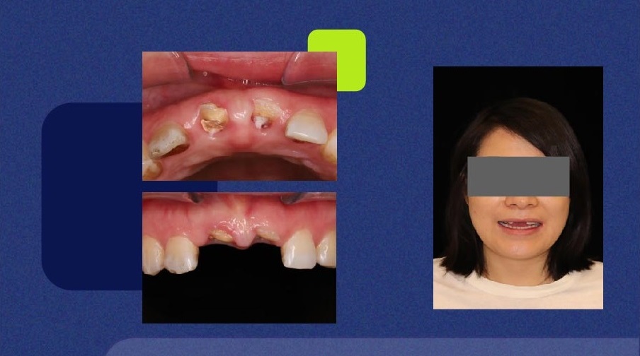
On February 19, 2021, Ms. Li broke her anterior teeth due to trauma. She felt that the aesthetics and function were seriously affected, and she went to the clinic to repair her teeth.
Oral examination:
*There is no defect in the lip, the opening degree is normal, and there is no snapping in the joint area.
*A1, B1 tooth root can be seen in the mouth
*Superficial overbite and overburden of anterior teeth, slightly lower frenulum position
*The overall mouth hygiene is slightly worse, with more dental calculus, soft scale and pigmentation.
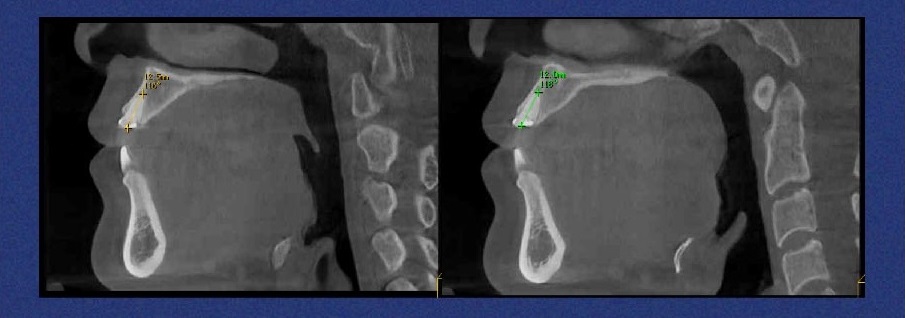
*CT showed that A1, B1 root length was about 12MM, alveolar width>7MM, no obvious abnormal periodontal
CT Images:
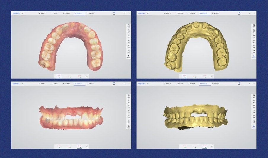
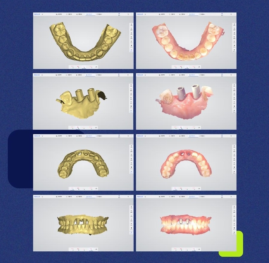
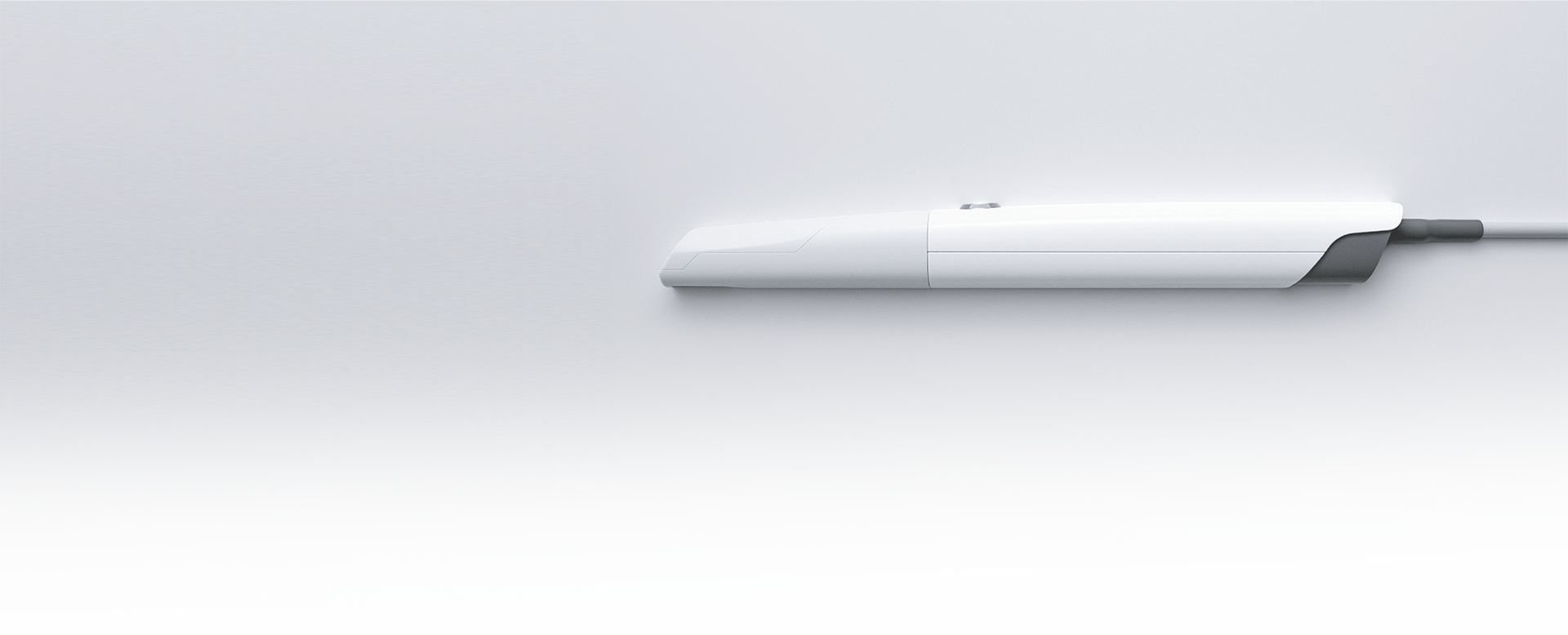
PANDA P2 Scanning:
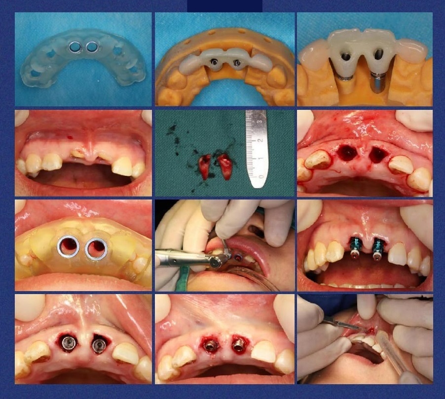
After communication, the patient chooses to immediately extract, implant and repair.
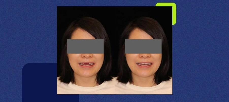
Preoperative DSD Design
Implant Surgery Photos
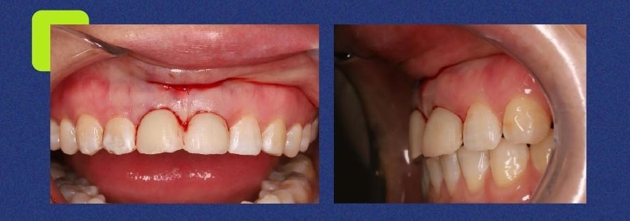
Intraoral Photo After Surgery
CT Images After Dental Implant
Phase II Restoration of PANDA P2 Scanning Data
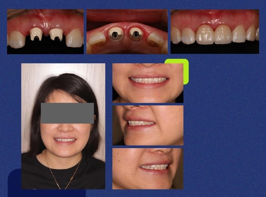
On July 2, 2021, the patient finished wearing the teeth
The whole process is digitally designed to complete the production, and the patient’s oral conditions are accurately replicated through PANDA P2, combined with CT data to complete a complete set of surgical plans for soft and hard tissues.
Recent posts
Categories
- Case Sharing (1)
-
Product Introduction (16)
- Upgrade Your Scanning Experience with the New Panda Center
- Panda Scanner Orthodontic Simulation Intelligent Upgrade
- Panda Center New Features
- 2023.06.16 Freqty Cloud Update Log
- PANDA P3 New Functions Introduction
- PANDA P3 Intraoral Scanner will be Officially Launched on August 18th
- Why is the Digital Impression System Highly Recommended in Dentistry?
- New Function, New Inspiration
- How Can Intraoral Scanners Help with Orthodontics
- One-Click Installer is Now Available to All Customers
- Freqty Cloud Adds 3D Preview Function
- Freqty Cloud Adds a New Function
- Panda (Freqty) Cloud URL Link Change Notification
- Top Reasons Dentists Should Turn to Intraoral Scanner
- Panda Scanner — Focus on Intraoral Scanners
- Panda P2 Intraoral Scanner Opens A New Era of Oral Digitalization
-
Dental Exhibition (34)
- PANDA NEWS: DenTech China 2024 Ended Successfully
- MIDEC 2023 Malaysia Successfully Concluded!
- Panda Scanner Invites you to Participate in the MIDEC Malaysia 2023
- PANDA Series of Intraoral Scanners Were Well Received at IDEX 2023
- Day 1 of IDEX 2023 was a huge success for Panda Scanner!
- Panda Scanner Invites You to Participate in IDEX Istanbul 2023
- Panda Scanner Showcased PANDA smart Intraoral Scanner at IDS
- Great First Day of IDS 2023
- CDS 2023 was a Great Success
- The 28th South Dental China International Expo Ended Successfully
- AEEDC Dubai 2023 Ended Successfully
- Freqty Presents PANDA P3 Intraoral Scanner at AEEDC 2023
- Day 2 of AEEDC 2023 Dubai is Underway
- Panda Scanner at the 40th CIOSP
- Panda Scanner Sincerely Invites You to Attend AEEDC 2023
- Greater New York Dental Meeting 2022 Ended Successfully
- Panda Scanner is Waiting for You at Greater New York Dental Meeting
- GNYDM 2022 Invitation
- Panda Scanner will be at the GNYDM 2022 at the end of this month
- Panda Scanner is going to New York!!!
- IDEM 2022 in Singapore Ended Successfully
- The 22nd Brazilian Orthodontic Congress
- Panda Scanner is about to Participate in IDEM 2022, Singapore
- China Northeast Dental Exhibition Witnesses PANDA P2 Hard Power
- 39° CIOSP Exhibition Ended Successfully
- Collection of Exhibitions and Events from March to June 2022
- The 13th Congresso ABOR 2022 is in Fortaleza
- Panda Scanner Participates in AAO Annual Session
- Five Dental Exhibitions are Underway
- Panda Scanner Participates in the 2021 CDS Shanghai Dental Show
- PANDA P2 Intraoral Scanner at 2021 IDS Cologne International Dental Show
- Sino-Dental x PANDA P2 Intraoral Scanner
- PANDA P2 appeared at the South China International Dental Show
- The 27th South China International Dental Exhibition
- Cooperation Case (5)
-
Training Courses (12)
- PANDA ACADEMY: How to do a 360° denture scan?
- PANDA ACADEMY: Step-by-step Scanning of Edentulous Cases
- PANDA ACADEMY: How to Scan an Implant Case?
- PANDA ACADEMY: Precautions for Wreless Connection of PANDA P4
- PANDA ACADEMY: How to Scan the Scanbody?
- PANDA ACADEMY: How to scan edentulous cases?
- PANDA ACADEMY: What can an intraoral scanner bring you?
- PANDA ACADEMY: How to Activate Your Scanner?
- PANDA ACADEMY: How to Connect Your Scanner?
- PANDA ACADEMY: Follow the digital dentistry trend
- PANDA ACADEMY: Recommended Computer Configuration
- Together with One Heart to Build a Better Future Intraoral Scanner Data Docking and Application Training Class
-
Health Tips (6)
- How Digital Dentistry Can Make Dentistry More Effective
- How Intraoral Scanners Help Dental Laboratories?
- How Important are Dental Intraoral Scanners?
- Are Intraoral Scanners Beneficial for Your Practice?
- How to Make Better Use of Your Intraoral Scanner
- Top 6 Tips for Choosing the Right Intraoral Scanner
-
Activity (13)
- Participate in the online lucky draw to win a free PANDA P3 intraoral scanner!
- Get Your Free Intraoral Scanner at AEEDC 2024
- Get Your Free Intraoral Scanner at AEEDC 2024
- Greater New York Dental Meeting 2023 Ended Successfully
- Coming Soon in November!
- Freqty Public Welfare Trip
- National Day Holiday Notice
- Panda Scanner Scan Speed Competition
- Digital Application Experience Activity and the First Intraoral Scanner Skills Competition
- PANDA P3 Officially Launched
- PANDA P2 Intraoral Scanner Settled in the Oral Health Science and Technology Museum
- Oral Medical Industry Association Annual Meeting ended successfully
- 2021 Customer Thanksgiving Event


